Monday, August 5, 2024

How much oxygen can a gram of haemoglobin carry?


3,740,000,000,000,000,000,000


If all haemoglobin could carry oxygen then one gram of haemoglobin could carry 1.39 ml of oxygen:

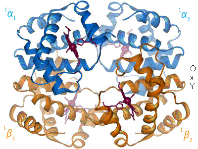
One molecule of haemoglobin will bind four molecules of oxygen

One 'mole' of haemoglobin will bind four moles of oxygen.

One mole of haemoglobin weighs 64,458.5 grams, and so:

1/64.458.5 mole of haemoglobin (i.e. 1 gram of haemoglobin) will bind 4 x 1/64,458.5 moles of oxygen

One mole of oxygen takes up 22.4 litres

4 x 1/64,485.5 x 22.4 = 1.39 ml of oxygen


But, not all haemoglobin is functional - some is damaged (dyshaemoglobin), and some likely to be combined with carbon monoxide, and neither of these can carry oxygen. When the actual oxygen carrying capacity of haemoglobin is measured various values have been found, but 1.34 ml is often taken as the 'true' value, and is known as Hufner's number (or constant).


(BTW 1.39 ml of oxygen = 3,740,000,000,000,000,000,000 molecules of oxygen)

(Sorry, not really a picture! 😅)


Sunday, August 4, 2024

The oxyhaemoglobin dissociation curve can move!


The oxyhaemoglobin dissociation curve shows the relationship between oxygen saturation of haemoglobin and the PO2. Normal is shown in blue.

This relationship changes with several factors, as shown in the picture. A right shift means the haemoglobin holds the oxygen less tightly, and left shift means it will hold oxygen more tightly.

A practical physiological result of this is that in an exercising muscle (so raised temperature, lots of CO2, and a lower pH), haemoglobin will hold less tightly to oxygen - it will let it go exactly where it's needed.

When the haemoglobin gets back to the lungs (where CO2 drops because it's being exhaled, and so pH rises, and temperature drops a bit), then haemoglobin binds oxygen more tightly and efficiently takes it up - ready to deliver in the muscle.


Saturday, August 3, 2024

The oxyhaemoglobin dissociation curve


This is a rather rough and ready picture of the oxyhaemoglobin dissociation curve, but hopefully it shows the key points Ok. 

The y-axis is oxygen saturation of the haemoglobin, and the x-axis is partial pressure of oxygen around the haemoglobin.

It starts off from the origin with a gentle upward curve . At this point the haemoglobin is in the tense configuration and not binding oxygen as well as when it's in relaxed.

Then the curve gets steeper. As more oxygen binds to the haemoglobin it starts to move towards the relaxed state, and so binds oxygen more easily. This means that only a small increase in PO2 is needed for the haemoglobin to bind lots more oxygen - i.e. for the oxygen saturation to increase quite a lot.

Finally it gets flatter as almost all the haemoglobin binding sites have oxygen bound to them (it will obviously never get above 100%)


Haemoglobin, and oxygen binding


A haemoglobin molecule can 'bend' between two slightly different shapes known as the 'tense' and 'relaxed' conformations - shown on the left and right above respectively. The important point here is that the relaxed conformation can bind oxygen more easily.

Several factors will determine which shape a haemoglobin molecule takes, such as the pH, temperature and PCO2 of its surroundings. 

How much oxygen it has bound to it will also affect its shape, the more oxygen bound to it, the more it takes on the relaxed conformation and so the more easily it will bind more oxygen. This effect is what causes the shape of the oxyhaemoglobin dissociation curve - more on this later.

Image by BerserkerBen from Wikipedia under licence https://creativecommons.org/licenses/by-sa/3.0/ and split from the animated gif into two images.


Thursday, August 1, 2024

The Frank-Starling Law


The Frank-Starling law is fundamental to critical care. It describes the relationship between cardiac filling and cardiac contraction - essentially more filling = more contraction.

In normal physiology it means that output from the left heart matches output from the right heart - over a period of a few beats.

In critical care, it means that if we give a fluid-responsive patient fluid, then their cardiac output will increase.

The diagram above is plotted the opposite way round to the way it's generally drawn these days, and shows cardiac output on the x-axis, and filling pressure on the y-axis. It's from Patterson and Starling (1914) and shows the cardiac output from a number of unfortunate dogs as they varied the cardiac filling pressure. The key point being that as filling pressure increased so did output, though this fell off at a certain point. Equally important though is the considerable variation between different dogs. Although this was an experimental preparation the principle applies to humans in ICU - a particular filling pressure (i.e. CVP) does not mean a particular cardiac output.

Patterson SW, Starling EH (1914) On the mechanical factors which determine the output of the ventricles. J Physiol 48:357 – 379. Available from: https://doi.org/10.1113/jphysiol.1914.sp001669 (accessed 01.08.2024)


Wednesday, July 31, 2024

Haemoglobin


This is an image of a haemoglobin molecule using a ribbon diagram

Haemoglobin molecules have four subunits, normally these are two 'alpha' amino acid chains and two 'beta' amino acid chains - labeled as ∝1, ∝2, êžµ1, êžµ2.

Each subunit contains a haem group (the balls and sticks you can see in the diagram above). An oxygen molecule can reversibly bind to the haem group. So one haemoglobin molecule can bind four oxygen molecules, and no more.

A key point here is that it's not a rigid, immovable structure, and you can see it flexing between two conformations (more on this later).

Image by BerserkerBen and used unmodified from Wikipedia under licence https://creativecommons.org/licenses/by-sa/3.0/

How much oxygen can a gram of haemoglobin carry?

  3,740,000,000,000,000,000,000 If all haemoglobin could carry oxygen then one gram of haemoglobin could carry 1.39 ml of oxygen: One molecu...